The transformation after viral infection
- Cristina Howard-Varona
- Sep 12, 2023
- 2 min read

Updated: Sep 23, 2023
Behind the scenes of the 2020 publication that can be found here.
When a cell is infected with a virus it becomes a new entity. This new entity, termed “virocell”, has a new metabolism and behavior: it no longer works to grow and multiply the cell; it deviates all energy and resources towards multiplying the virus. But, what happens if the same cell is infected with different viruses? Are the virocells the same?
We answered this question in our publication, “Phage-specific metabolic reprogramming of virocells”. There, we at the Ohio State University and University of Michigan teamed up with various national laboratories (Joint Genome Institute, Pacific Northwest National Laboratories, and the Environmental Molecular Sciences Laboratory), through a User Award, to design and execute an experiment where the same bacterial cell was independently infected with two very different viruses (phages). Then, we monitored through time those infections, collecting samples that would enable us to “see” what was happening inside the cells throughout the stages of infection, from the moment that the phage encountered the cell until it released all of its progeny through cell lysis. This was not an easy task. We collected samples that monitored how cells were dying and how phages were reproducing. Then, to study what processes were happening inside those virocells, we performed RNA-sequencing, a technology that enables us to simultaneously visualize changes in expression of all phage and cellular genes. Because DNA and proteins are also important components of an organism, we also followed DNA replication and degradation with PCR and microscopy, as well as changes in the proteins, through mass-spectrometry of all phage and cellular proteins. Finally, to see how the metabolisms were changing, we performed metabolomics, which captured small molecules such as sugars or lipids. All of these samples together provided us with a clear picture of what was happening inside our phage-infected cells, at every stage of infection. But, they were not easy to collect. Only thanks to an “army” of undergraduate and graduate students from both academic institutions, technical staff, myself and the two professors in the project all working together, we were successful.
Three years took to publish the work, during which time we had a lot of fun discovering new science. We discovered that when the same cell is infected with different viruses, it creates drastically different virocells. Each virocell behaves differently depending on the needs of the virus, and the needs of the virus depend on how genetically similar that virus is to the cell. If the virus shares many genomic features, such as the ability to easily produce proteins with cellular machinery, the virocell will work less hard at reproducing the virus, because it can readily use the resources that the cell provides. Instead, if the viral genetic information is very dissimilar to that of the cell that it infects, then it has to work hard at deviating energy and resources towards building the components of the new viral progeny. This science provides an exciting window into predicting the multiple lives that a cell may have, depending on the genetic composition of the cell and the infecting virus.
You can read the science communication piece released by the DOE here.
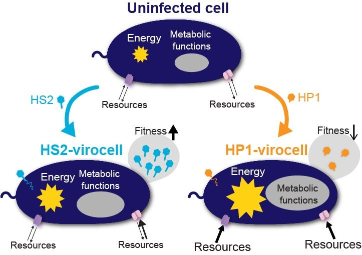
The same cell, when infected with two different viruses, is transformed into two very different virocells.
Comments